
3. Oesophagus 4. Stomach
5. Small Intestine 6. Large Intestine
(a) Mouth Cavity: Mouth is bounded by the immovable upper jaw and movable jaw. It is formed by cheeks and hard and soft palates. Hard palates forms the roof of the mouth and continues posteriorly into soft palate. Hard palate bears transverse ridges, the rugae. The hinder free part of the soft palate freely hangs down as a small flap called uvula.
 A muscular tongue lies on the floor of buccal cavity. The rough surface on the tongue are called papillae. Taste buds are located on the tongue which contain receptors sensitive to sweet, salty, sour and bitter substances. Mouth bears three pairs of salivary glands which secrets into the mouth cavity.
Teeth: Human teeth are thecodont, diphyodont and heterodont. They are found embedded in jaw sockets (thecodont). Humans have two sets of teeth. The milk teeth appear first, and are progressively replaced by the permanent teeth (diphypodont). Human teeth have different shapes and sizes and possess uneven biting surfaces. Human possesses up to 3d2 permanent teeth, consisting of 8 incisors (i), 4 cannines (c), 8 premolars (pm) and up to 12 molars (m).

The arrangement of teeth can be conveniently expressed in the form of a dental formula. Human permanent dental formula is:
I - 2/2, C - 1/1, PM - 2/2, M - 3/3 = 32
Third molar appears only after the age of 20 years. So, they are called wisdom teeth. But sometimes, they may not appear in certain person. The number, size and shape of teeth are related to diet. on the basis of structure and function of each type of tooth, four different of teeth (heterodont) are as follows:
- Incisors- They are situated at the front of buccal cavity. They have flat and sharp edges which are used for cutting and biting food.
- Canines- They are pointed dagger-shaped teeth. They are poorly developed in human, but highly developed in carnivores used for tearing flesh.
- Premolars- They are broad and strong having one or two roots and two cups, (projections on the surface of a teeth). They are specialized for crushing and grinding food, although in human they may also be used to tear food.
- Molars- They have more than one root; upper molars have three roots, lower molars have two. Each has four to five cups. They are used to crush and grind.

A typical tooth consists of three parts.
(i) Crown (ii) Neck (iii) Root
(i) Crown : It is the exposed part of tooth. It is covered with the hardest substance called enamel. It is relatively resistant to decay. Internally, it has hard substance called dentine.
(ii) Neck : It is a short part embedded in gums.
(iii) Root : This part is embedded in body jaw that holds the tooth. Internally, root has pulp cavity which contains of nerve fibers, odontoblasts ( dentine-producing cells), blood vessels, which deliver nutrients to the living tissues of the tooth . The root of the tooth is covered with cement, a hard substance similar to bone.
2. Pharynx : Pharynx is situated behind the soft palate. The pharynx connects with six openings- two internal nares from the nasal cavity, the two eustachian tube leading to the middle ears, the glottis opening into the larynx and the esophagus leading to the stomach.
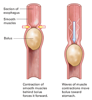
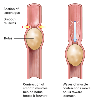
3. Esophagus: Esophagus or foods pipe is a long (about 25cm) narrow muscular tube leading directly to the stomach piercing the diaphragm. It quickly conveys food (bolus) and fluids by peristalsis from pharynx to the stomach.
4. Stomach: Stomach, the widest organ, is large, curved and J-shaped elastic sac situated just below the diaphragm on the left side of abdominal cavity. It has three parts.
(i) Cardiac part- It is upper part of stomach, near the heart. Esophagus opens into it.
(ii) Pyloric part- It is lower part nearest to the duodenum.
(iii) Fundic part- It is middle part of stomach.

5. Small intestine: It is very long coiled tube of about 6.5m. It is divided into three parts-
(i) Duodenum: Duodenum is ‘U’ shaped curved about 25 cm long. It receives the opening of the common bile duct, formed by the union of the bile duct and the pancreatic duct. It is the main part for the digestion of food . Internally, its wall contains numerous glands, which secrete the intestinal juice containing several digestive enzymes.
(ii) Jejunum: It is the middle part of the small intestine, narrower than duodenum. It is 2.5 m long and coiled.
(iii) Ileum: It is highly coiled part. Its inner surface possesses with numerous small finger projections called villi, which greatly increase the surface area for absorption. It is about 3.5 m long.
Large intestine : Large intestine is shorter but wider than small intestine. It measures about 1.5 meter long. It is mainly responsible for the absorption of water and discharging undigested food particles. It is divide into three parts (i) Caecum (ii) Colon (iii) Rectum


(i) Caecum: The caecum is a pouch like structure which is about 6 cm. the junction guarded by the ileocaecal valve. It prevents the back flow of food into small intestine. Attached to the caecum is the slender vermiform appendix which is 8-10 cm long. It is thought to be vestigial in man but functional only in herbivores.
(ii) Colon: It looks like an inverted U tube which is divided into four regions- ascending colon, transverse colon, descending colon and sigmoid colon. Its walls have constricted pouch like structures called haustra. The opening of caecum and colon is guarded by an ilio-caecal sphincters.
Digestive Glands
Digestive glands associated with digestion of food are-
(a) Salivary glands (b) Gastric glands (c) Liver
(d) Pancreas (e) Intestinal glands
(a)Salivary glands: These glands secrete saliva. Three types of salivary glands located into mouth cavity-
(i) Patroid glands- located at the base of ear,
(ii) Sub-maxillary glands- located posterior of mouth floor,
(iii) Sub-lingual glands- located below the tongue.
Saliva : Saliva is a viscous colorless, cloudy and opalescent fluid. Its average pH value is 6.8. It contains 98.5 to 99% water and 1 to 1.5% of a dense residue. It has an enzyme called salivary amylase or ptyalin used for digestion of starch. A normal person secretes 1.0 – 1.5 liter of saliva daily.
Function of saliva: saliva performs a number of functions . These are
· It possesses an enzymes called ptyalin for digestion of starch.
· It moistens dry food to facilitate easy swallowing.
· It dissolves the soluble substances such as sugar and salt.
· It keeps the mouth and teeth clean.
· It makes the food delicious to taste.
· It stimulates the taste buds.
· It contains three buffering systems; bicarbonate, phosphate and mucin.
(b)Gastric glands : The wall of stomach has numerous deeply pitted gastric glands, which secrete gastric juice. A normal person secretes about 2-3 liters of gastric juice daily. The gastric juice is light colored, thin and transparent fluid. Three types of gastric glands are.
(i)Parietal (oxyntic) cells – secrete HCL,
(ii) Chief or peptic (Zymogen) cells- secrete pepsin and other enzymes,
(iii) Mucous (Goblet) cells- secrete mucin.
Composition of gastric juice: It contains about 90% water,0.5% HCL and rest is mineral such as potassium chloride and phosphates, mucin and enzymes like pepsin, rennin and gastric lipase.
Functions of HCL
· It changes the pH of food to acidic.
· It destroys bacteria or microbes present in food.
· It activates the inactive pepsinogen to active pepsin.
· It digests the hard substances like bones.
· It controls the opening and closing of pyloric aperture.
Functions of pepsin: Initially pepsin is released in an inactive form i.e. pepsinogen which is activated by HCL into active pepsin. The active pepsin hydrolyzes the proteins into proteoses and peptones.
Thanks for sharing the valuable information here. So i think i got some useful information with this content. Thank you and please keep update like this informative details.
ReplyDeletePainless Dental Treatment In Chennai
Best Dental Clinic In Adyar